

Extraordinary MRI images showing what happens inside the human body during sexual intercourse have resurfaced online, leaving people fascinated by the unusual scientific experiment behind them.
The images were captured during groundbreaking research that began in the Netherlands in 1991, when a couple volunteered to have sex inside a magnetic resonance imaging scanner.
Although the experiment took place more than three decades ago, its findings challenged longstanding assumptions about human anatomy and eventually became the subject of a widely discussed medical study.
The research was published in the British Medical Journal in 1999 and has continued to attract attention ever since.
Now, as the images circulate again on social media, people are discovering what the researchers observed and why the findings were considered so significant.
What did the MRI images reveal about sex?
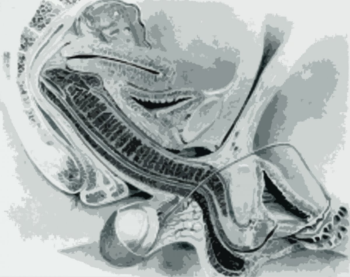
The most surprising finding involved the shape and positioning of the penis and vagina during penetrative intercourse.
For centuries, anatomical illustrations had often depicted the vaginal canal as a relatively straight passage, with the penis entering along a similarly straight path.
However, the MRI scans showed that the penis can take on a curved, boomerang-like shape during intercourse, adapting to the positioning and contours of the surrounding anatomy.
The researchers also observed that the vaginal canal and surrounding structures did not follow the simple straight-line arrangement suggested by some earlier illustrations.
Instead, the images revealed a more complex relationship between the two bodies, including how their internal structures were positioned during penetration.
The findings offered researchers a rare opportunity to examine sexual anatomy directly rather than relying entirely on diagrams, external observations or theoretical models.
Importantly, the curvature visible in the scans did not mean the penis was being injured or unnaturally bent. It reflected its position within the anatomy during the particular circumstances of the experiment.
The images helped demonstrate that human anatomy during intercourse is more complex than traditional simplified drawings might suggest.
How did scientists manage to capture the images?
The unusual research began with Dutch scientist Menko Victor van Andel, commonly known as Pek, who wanted to investigate human sexual anatomy using MRI technology.
At the time, MRI scanning was already widely used to examine internal organs and tissues, but capturing images during sexual intercourse presented obvious practical difficulties.
For one thing, the participants needed to fit inside the scanner's narrow opening.
They also had to remain sufficiently still for researchers to obtain usable images.
Unlike a conventional photograph, an MRI scan requires specialised equipment and careful positioning to produce a clear picture of the body's internal structures.
These challenges made the experiment far more complicated than simply asking two people to lie inside a machine.
Nevertheless, van Andel eventually found two willing volunteers.
The couple who volunteered for the unusual experiment
Ida Sabelis and her partner Jupp agreed to participate in the first experiment in 1991.
Ida was acquainted with the scientist through a personal connection and later explained that she had agreed to help partly because she wanted to contribute to a better scientific understanding of women's bodies.
She also had an interest in challenging traditional assumptions about female anatomy.
Despite the scientific purpose of the experiment, the couple found themselves dealing with a rather unusual situation.
They needed to position themselves together inside the confined space of an MRI scanner while researchers prepared to capture images.
Ida later recalled that there was considerable laughter as they attempted to get comfortable.
Originally, the experiment was expected to involve the missionary position.
However, Ida suggested that a side-lying position would be more practical because of the limited space available.
The adjustment helped the couple fit inside the scanner and allowed the researchers to proceed.
What happened inside the MRI scanner?
Once positioned inside the machine, Ida and Jupp were asked to engage in sexual intercourse while the researchers prepared the equipment.
They then had to stop moving and remain still at specific moments so that the scanner could capture images.
This created an unusual combination of intimacy and scientific observation.
Unlike modern video recordings, the resulting MRI images provided detailed views of internal anatomy rather than conventional footage of the participants.
The scans allowed researchers to examine the positioning of reproductive structures during intercourse.
When Ida later saw the images, she described being struck by the way the two bodies fitted together.
The experience provided an entirely different perspective on an activity that is familiar to many people but rarely observed from inside the body.
Why did the discovery surprise researchers?
Historically, scientific understanding of sexual anatomy had been influenced by anatomical drawings and observations that could not directly show the positioning of internal structures during intercourse.
Some traditional illustrations presented penetration as a relatively straightforward process involving two nearly straight structures.
The MRI images challenged that simplified representation.
They showed how the penis could curve during penetration and how the surrounding female anatomy accommodated its position.
This was particularly interesting because the findings illustrated anatomical relationships that had previously been difficult to document.
The experiment did not establish that every couple's anatomy behaves identically during sex.
Individual anatomy, sexual position and other factors can influence the precise arrangement of structures.
However, the images provided a useful demonstration of why static diagrams do not always capture the body's behaviour during real activities.
The research was expanded into a formal study
Following the initial experiment, van Andel and his colleagues continued investigating sexual anatomy using MRI scanning.
Between 1991 and 1999, additional volunteers participated in research designed to capture further images.
The study involved consenting adults who were informed about the procedures and could withdraw from participation.
Researchers attempted to record intercourse in different circumstances, including the missionary position.
However, the unusual environment made the experiment challenging for some participants.
The confined scanner, the presence of researchers and the need to remain still were hardly typical conditions for sexual activity.
According to accounts of the research, some participants required medication to help maintain an erection.
Ida and Jupp were reportedly able to complete their original experiment without such assistance.
These practical difficulties were an important limitation of the study, since sexual activity inside an MRI scanner may not perfectly represent what happens in a more natural setting.
What did the 1999 British Medical Journal study conclude?
The research was published in the British Medical Journal in 1999 under the title Magnetic resonance imaging of male and female genitals during coitus and female sexual arousal.
It provided unusual visual evidence of the positioning of reproductive structures during intercourse.
The study described the boomerang-like configuration of the penis during penetration and examined the anatomy of the female reproductive organs in the captured images.
The researchers also used MRI scanning to investigate aspects of female sexual arousal.
Although the study was small and exploratory, it attracted attention because it demonstrated how medical imaging could be used to investigate questions that were difficult to study through traditional methods.
The findings were not intended to provide universal conclusions about every sexual position or every person's anatomy.
Instead, they contributed to the scientific description of human sexual physiology and demonstrated the potential value of imaging technology in this field.
Why did the study become so famous?
The unusual subject matter and striking images helped make the paper one of the British Medical Journal's most widely discussed publications.
Unlike many medical studies, which attract attention primarily within specialist scientific communities, this research appealed to a much broader audience.
People were fascinated by both the findings and the practical challenges involved in conducting the experiment.
The fact that two volunteers had managed to have intercourse inside an MRI scanner became almost as famous as the scientific observations themselves.
The paper continued to be read long after its original publication.
In 2019, the BMJ revisited the research to mark its twentieth anniversary, reflecting its lasting popularity.
Social media users react to the resurfaced images
More recently, the scans have circulated on platforms including TikTok, introducing the research to people who were not even born when the original experiment took place.
Some viewers have expressed surprise at the internal positioning shown in the images.
Others have been more interested in how the volunteers managed to fit inside the scanner.
The research has also prompted conversations about the history of medical knowledge and the importance of studying female anatomy in greater detail.
Although the experiment may initially sound amusing, its purpose was to answer genuine scientific questions.
The resulting images demonstrated how direct observation can challenge assumptions that have been repeated for generations.
What can the research tell us about the human body?
The study remains an example of how medical imaging can reveal anatomical details that are difficult to understand through conventional illustrations alone.
Its central finding was that the positioning of reproductive structures during intercourse is more complex than a simple straight-line model suggests.
It also highlighted the importance of investigating female sexual anatomy using scientific methods rather than relying exclusively on historical descriptions.
However, the findings should be understood within the limitations of a small study conducted under unusual experimental conditions.
The images do not establish a single correct sexual position, nor do they imply that the same anatomical configuration occurs during every sexual encounter.
Nevertheless, more than three decades after the first experiment, the scans continue to fascinate people because they offer an extraordinary view of something usually hidden from sight.
What began as an unconventional scientific investigation has become a lasting example of how curiosity, willing volunteers and modern technology can produce surprising insights into the human body.
Sources: VT, December 30, 2024; van Andel and colleagues, British Medical Journal, 1999, "Magnetic resonance imaging of male and female genitals during coitus and female sexual arousal".